
Fingers are easily injured from everyday activities, and finger injuries are some of the most common traumatic injuries seen in an emergency room. Injuries may range from simple bruises or contusions to broken bones and dislocations of the joints. Understanding the basic anatomy of the hand and fingers is useful in understanding different types of finger injuries, broken fingers, and how some treatments differ from others.
 The hand is divided into three sections: wrist, palm, and fingers. There are eight bones in the wrist, which move together to allow the vast ranges of motion of the wrist. The palm, or mid-hand, is made up of the metacarpal bones. The metacarpal bones have muscular attachments and bridge the wrist to the individual fingers. These bones frequently are injured with direct trauma such as a crush from an object or most commonly from a punching injury.
The fingers are the most frequently injured part of the hand. Fingers are constructed of ligaments (strong supportive tissue connecting bone to bone), tendons (attachment tissue from muscle to bone), and three bones called phalanges. There are no muscles in the fingers. Fingers move by the pull of forearm muscles on the tendons.
- The three bones in each finger are arranged in the same manner. The finger bones are named in their relation to the hand. For example, the first bone is the proximal phalanx. The second bone is the middle phalanx. The smallest and farthest from the hand is the distal phalanx. The thumb is the shortest finger and does not have the middle phalanx.
- Knuckles on the back of the hand are joints formed by the bones of the fingers. They are commonly injured or dislocated with trauma to the hand. Each joint has a specific name depending on its location and the bones involved.
The first and largest knuckle is the junction between the palmar bones and the fingers. Medically, it is the joint of the metacarpals and phalanges. This joint commonly is injured in closed fist activities, and a fracture of this area is commonly known as a boxer’s fracture.
The next knuckle out toward the fingertip is the joint closest to the hand and between the finger bones. It is termed the proximal inter-phalangeal joint (PIP). This joint may be dislocated in sporting events when a ball or object directly strikes the finger.
The farthest joint of the finger is the distal inter-phalangeal joint (DIP). Injuries to this joint usually involve a fracture (a break) or tendon tearing (avulsion) injury.
Broken finger symptoms
 Broken fingers frequently cause immediate pain after trauma and sometimes a deformed finger either at a joint (commonly a dislocation) or through the bone as a fracture. If there is no deformity, a sharp pain usually is felt very specifically at the injury site.
Sometimes you are not sure if the finger is broken, and you try to bend the finger in question. A true fracture usually will be painful, but do not be fooled by a finger that has some range of motion and dull pain. Depending on their stability, some fractures may hurt more than others.
As time goes on, usually within the next 5-10 minutes, swelling and bruising of the finger will occur and the finger will become stiff to move. Swelling is not as specific as pain and therefore may affect the adjacent fingers as well.
If the fracture is severe, bruising from released blood may be seen immediately.
Finally, if the swelling is excessive, numbness of the finger may occur because the nerves in the fingers are compressed.
When to Seek Medical Care
The doctor will need an x-ray to evaluate the position of the broken finger bones. The best medical treatment would be found in an urgent care facility or a hospital’s emergency department, which have the needed supplies for x-ray evaluation and splinting.
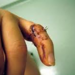
Rarely, a surgical procedure is needed to stabilize the fracture. Complications of this may include infection. Signs of infection are fever, increasing redness, swelling, severe pain of the finger, or even discharge of pus and a foul smell from the surgery site. If these symptoms occur, go to the emergency department or your surgeon immediately to be evaluated.
Exams and Tests
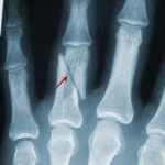
X ray of broken finger
The mainstay of diagnosing finger fractures is an x-ray. Temporary splinting, ice, and pain control are helpful supportive treatments. The type of fracture will determine the treatment. Each fracture pattern has specific characteristics that need to be addressed.
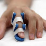
If there is a simple fracture, the doctor will splint the injured finger. The whole hand may be put at rest and splinted for comfort.
With more complex injuries, the doctor may seek the advice of an orthopedic (bone and joint specialist) or hand surgeon (someone with special training in hand surgery).