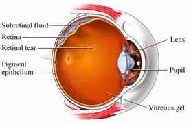
Treatment for a detached retina typically involves immediate surgery in order to repair the condition. The specific type of surgery your healthcare provider recommends will depend on the type, size, and location of the detached retina. The goals of surgery are:
- To reattach the retina.
- To prevent or reverse vision loss.
Laser Surgery and Cryopexy
 For small holes and tears, laser surgery or a freezing treatment called cryopexy may be recommended, which can usually prevent retinal detachment and preserve almost all vision. These procedures are usually performed in the doctor’s office.
Treating a retinal tear may be useful if the tear is likely to lead to detachment. Symptoms such as floaters or flashing lights are key factors in deciding whether to treat a tear. A tear that occurs right after a posterior vitreous detachment (PVD) with symptoms is usually much more dangerous and more likely to progress to a retinal detachment than one that occurs without symptoms.
In deciding when to treat a retinal tear, your doctor will evaluate whether the torn retina is likely to detach. If the tear is very likely to lead to detachment, treatment can usually repair it and prevent detachment and potential vision loss. If the tear is not likely to lead to detachment, you may not need treatment.
- Laser surgery (photocoagulation). During photocoagulation your surgeon directs a laser beam through a contact lens or ophthalmoscope designed for this procedure. The laser makes burns around the retinal tear, and the scarring that results usually “welds” the retina to the underlying tissue.
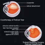
- Freezing (cryopexy). During cryopexy, your surgeon uses intense cold to freeze the retina around the retinal tear. After a local anesthetic numbs your eye, your surgeon applies a freezing probe to the outer surface of the eye directly over the retinal defect. This freezes the area around the hole, leaving a delicate scar that helps secure the retina to the eye wall.
After your procedure you’ll need to remain relatively still for the next two weeks or so, as the bonds created by your procedure strengthen.
Scleral Buckle, Vitrectomy, and Pneumopexy
 For some cases, other types of surgery for detached retina may be recommended. These procedures may be done in conjunction with photocoagulation or cryopexy. The type, size and location of the retinal detachment will determine which surgical approach your eye surgeon recommends. In general, these surgeries can successfully treat most cases of retinal detachment, although a second treatment is sometimes necessary. These surgeries include scleral buckling, vitrectomy, or pneumopexy.
Scleral Buckling
A procedure called scleral buckling involves suturing a piece of silicone rubber or sponge to the white of your eye (sclera) over the affected area. The silicone material indents the wall of the eye, relieving the tugging of the vitreous on the retina. When you have several tears or holes or an extensive detachment, your surgeon may create an encircling scleral buckle that goes around the entire circumference of your eye like a belt. The buckle usually remains in place for the rest of your life. A scleral buckle is a tiny synthetic band that is attached to the outside of the eyeball to gently push the wall of the eye against the detached retina.
Vitrectomy and Pneumopexy
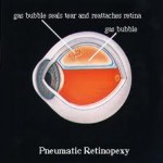
During a vitrectomy, the doctor makes a tiny incision in the sclera (the white of the eye). Next, a small instrument is placed into the eye to remove the vitreous, a gel-like substance that fills the center of the eye and helps the eye maintain a round shape. Then, gas is often injected into the eye to replace the vitreous and reattach the retina (known as a pneumopexy); the gas pushes the retina back against the wall of the eye. During the healing process, the eye makes fluid that gradually replaces the gas and fills the eye.
With no new fluid passing through the retinal tear, fluid that had previously collected under the retina is absorbed, and the retina is able to reattach itself to the back wall of your eye. Depending on where the retinal detachment is located in your eye, you may need to hold your head in a certain position for several hours in order to keep the bubble in place. With these two surgical procedures, after a couple of weeks, laser surgery or cryopexy is used to “weld” the retina back into place.
Expected Results of Surgery for Detached Retina
 With modern detached retina surgery, more than 90 percent of cases can be successfully treated. In some cases, however, a second surgery is needed.
The visual outcome is not always predictable following surgery. The final visual result may not be known for up to several months following surgery for detached retina. Even under the best of circumstances, and even after multiple attempts at repair, treatment sometimes fails, and vision may eventually be lost. Visual results are best if the retinal detachment is repaired before the macula (the center region of the retina responsible for fine, detailed vision) detaches.