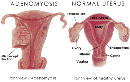
Author Archives: alwin

Adenomyosis of the uterus

Known as “Endometriosis of the uterus”, Adenomyosis is benign and does not cause cancer. Most commonly, the disease affects the back wall (posterior side) of the uterus. The endometrial cells penetrate deep into the uterine muscle (myometrium). When this occurs, the uterus is enlarged usually more than twice the normal size and very hard. The disease may be localized with well-defined borders or diffuse, meaning having no limits or borders. When this localized disease is found it is called adenomyoma. These adenomyomas can be located at different depths of the uterine muscle and can penetrate into the uterine cavity, becoming submucosal tumors. Continue reading

Adjuvant chemotherapy for lung cancer

The use of chemotherapy after curative resection is standard in many common malignancies such as breast and colon cancer and is commonly described as adjuvant therapy. The magnitude of benefit with respect to improvement in disease-free and overall survival in many cases is often small in absolute terms especially in the case of breast cancer where absolute potential improvements may only be in the range of 5% at 10 years. Lung cancer is the most lethal of the common malignancies with non-small cell lung cancer (NSCLC) comprising approximately 80% of cases. Unfortunately, only a minority of patients present with resectable disease, and despite this, the median survival with curative intent surgery is relatively poor (pathological stage I disease 67% at 5 years, pathological stage 2b disease 39% at 5 years). NSCLC is predominantly a disease of the elderly with median age at diagnosis of 68 years, and its management is often complicated by the relatively high rate of concomitant illnesses. Continue reading

Adrenal cancer Part 4

Radiation Therapy

Radiation therapy is used in a number of cancers as both the main method of killing cancer cells or in combination with surgery (either before or after). The radiation comes in the form of high-energy x-rays that are delivered to the patient only in the areas at highest risk for cancer. These x-rays are similar to those used for diagnostic x-rays, only of a much higher energy. The high-energy of x-rays in radiation therapy results in damage to the DNA of cells, causing tumor cells to die. Continue reading

Adrenal cancer Part 3

How are adrenal cortical cancers staged?

In addition to diagnosing adrenal cortical cancers, the radiographic imaging performed also helps to determine the stage of the patient. According to the seventh edition of the American Joint Committee on Cancer (AJCC), which defines cancer staging systems, patients with adrenal cortical cancer are divided into one of four stages. Please note that there is no AJCC staging system for medullary adrenal carcinoma. Continue reading

Adrenal cancer Part 2

What causes adrenal cortical cancers and am I at risk?

Each year, there are approximately 500 cases of adrenal cortical cancers in the United States. These most commonly occur in patients between the ages of 30 and 50; however, children under the age of 5 develop adrenal cortical cancers at a higher rate than the rest of the population. Males are more likely to develop non-functioning adrenal carcinomas, while females are more likely to develop functioning adrenal carcinomas. In general, causes of adrenal cortical cancers are unknown. They are not associated with smoking, and do not run in families. Despite this, certain rare genetic syndromes such as Beckwith-Wiedemann syndrome, and multiple endocrine neoplasia, have been associated with adrenal cortical cancers. Continue reading

Adrenal cancer Part 1

What are the adrenal glands?

The adrenal glands are small glands that are located just above each kidney (they are sometimes called the suprarenal glands for that reason). The adrenal glands are surrounded by a layer of connective tissue and a layer of fat. They are triangular in shape and consist of several distinct parts: Continue reading

Adenosine thallium scan

Adenosine thallium scan: A method of examining the heart to obtain information about the blood supply to the heart muscle. Special cameras take a series of pictures of the heart. Radioactive thallium is injected into the bloodstream and serves as a tracer. The tracer attaches to certain cells and makes them visible to the special camera. The tracer attaches to the muscle cells of the heart so the imaging camera can take pictures of the heart muscles. If an area of the heart does not receive an adequate flow of blood, the cells in the underserved area do not receive as much tracer and it appears as a darker area on the picture taken by the camera. Continue reading

Acute hepatitis B Part 2

What determines the outcome of acute hepatitis B?

The body’s immune response is the major determinant of the outcome in acute hepatitis B. Individuals who develop a strong immune response to the infection are more likely to clear the virus and recover. However, these patients also are more likely to develop more severe liver injury and symptoms due to the strong immune response that is trying to eliminate the virus. On the other hand, a weaker immune response results in less liver injury and fewer symptoms but a higher risk of developing chronic hepatitis B. People who recover and eliminate the virus will develop life-long immunity, that is, protection from subsequent infection from hepatitis B. Continue reading

Acute hepatitis B Part 1

What is hepatitis?

The term ‘hepatitis’ simply means inflammation of the liver. Hepatitis may be caused by a virus or a toxin such as alcohol. Other viruses that can cause injury to liver cells include the hepatitis A and hepatitis C viruses. These viruses are not related to each other or to hepatitis B virus and differ in their structure, the ways they are spread among individuals, the severity of symptoms they can cause, the way they are treated, and the outcome of the infection. Continue reading